
A massive advancement in personalised medicine and the future of cancer treatment has and is happening at the PA Hospital campus in Brisbane.

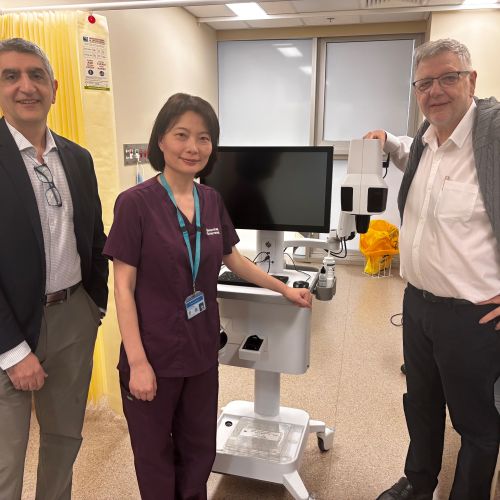
Based at the Translational Research Institute on the hospital campus, Dr Arutha Kulasinghe in collaboration with Akoya Biosciences has been able to use spatial profiling technology to give the most detailed look at tumours yet and his discovery has amazed the medical world.
Dr Kulasinghe is confident the imaging technology, which is akin to the ‘google maps’ for cancer, will at some stage in the future be key in advising clinicians on the likelihood of immunotherapy being successful in treating a patient’s cancer.
The technology so far shows the most promise in head and neck cancers and lung cancer but can easily be adapted to explore cell and tissue interactions in numerous other forms of solid cancers.
The progress means the research is a step closer to being able to be proven through a clinical trial, so that the exciting leap forward in guiding clinicians on the most effective treatments can benefit cancer patients across the world.
“Ultimately, we want a standard test that a pathology lab can do to predict responses to immunotherapy, and we are a way off doing that, but I think we will make inroads into that space because of this technology and this advancement,” Dr Kulasinghe said.
“While chemotherapy works to kill cancer cells, immunotherapy aims to boost the body’s own immune system to fight cancer. It has shown great promise in a subset of head and neck cancer, and lung cancer patients leading to long term benefits and better quality of life.”
“Immunotherapy is a pillar of cancer therapeutics, but what we really need to understand is which patients are most likely to respond and then give the therapies to those patients. In doing so, you reduce the chance of an ineffective therapy and possible immune related adverse effects, and you save the taxpayer hundreds of thousands of dollars, as these therapies are quite cost-intensive.
“This tissue atlasing approach is potentially transformative in that regard.”
Dr Kulasinghe said the exciting progression in their research started through their ongoing lab research but working with a spatial biology company brought it to the next level.
“We had successfully developed multi-colour staining protocols for head and neck cancers in the lab, however, in collaboration with Akoya Biosciences R&D team, we were able to develop this across an entire patient’s surgical specimen at single-cell resolution. This can give unprecedented insights into the tumour-immune cell biology, and the cellular architecture of the sample.
“We can now toggle through over 100 different biomarkers as we simultaneously scan the tissue section and understand the tumour-immune cell interactions, cellular architecture and the different cell populations ‘recognising’ or being ‘masked’ from the tumour. These are very valuable assessments when determining how the body’s immune system is recognising the tumour.
“In this patient’s tumour specifically, we could identify tertiary lymphoid structures easily, these are B cell rich structures and infiltrate the tumour, and can sustain an immune-responsive microenvironment – patients with these tend to be responsive to immunotherapy, and we can see this more clearly than ever now.
“This technology will show us that only certain regions of tumour would be likely to respond to immune checkpoint inhibitor therapy. We have never had imagery this detailed before and it is helping us to understand why patients may not fully respond to immune checkpoint inhibitor therapy which is the main form of immunotherapy.
“These therapies work in 15 to 40 per cent of head and neck cancers and lung cancers.
“Over half the population is not going to respond to these therapies, which cost about $150,0000 per patient per year, they can’t be given in a blanket approach because it is expensive, and some patients that don’t respond to immunotherapy can also have adverse effects, so it’s important to be able to predict which patients are going to respond.
“We think this tissue mapping approach will give us deep insights into why patients are responding or not responding, and we will be able to demonstrate that as well.”


The significant advancement in spatial profiling would not have been possible without the support of the PA Hospital and the PA Research Foundation and its supporters.
“This type of research is often considered high risk and funding can be challenging, but I think that we are now learning the benefits of funding this type of research. “
“We are eternally grateful to the Foundation and its donors.”